

Nanoartography 2022
Image credit: Pias K. Biswas, Mark Woollam, NanoArtography 2022
2022 Award Winners
Click on the images to see them in full size.
NanoArtography 2022 Numbers
Total submissions: 210 from 29 countries


FIRST PLACE
Bee Eco-Friendly
Yamini Mittal,
CSIR-Institute of Minerals and Materials Technology, Bhubaneswar, India
A beehive-inspired water filter! The scanning electron micrograph represents the transformation of Canna Indica plant waste into biochar through a combustion method. It shows porous beehive-like structure formation, exhibiting excellent wastewater treatment efficiency and significant bioelectricity generation when used as substrate in sustainable constructed wetland integrated microbial fuel cell (CW-MFC) technology. It reflects that “nature harmonizes creation and destruction, containing seeds of its own solution.” The image width is 0.01 mm.
SECOND PLACE (tie two ways)
The Tree of Life
Sharon Lim Xiaodai,
National University of Singapore, Singapore
Carbon atoms constitute up to ~ 50% of the dry mass of trees, and it is stored for life in wood products, just as this tiny tree is made up of beautifully arranged carbon atoms. This scanning electron microscopy image captures the water-assisted self-assembly of carbon nanotube micro-pillars (the tree) pre-constructed by running a focused laser beam through selected sites of an array of carbon nanotubes. When coupled with the interlacing nature of these carbon nanotube pillars, such a capillary-driven process makes an excellent sieve for nanoparticles. The image width is 0.063 mm.
SECOND PLACE (tie two ways)
Mount. MXene
Grace Cooksley,
University of Brighton, United Kingdom
This artwork was created by preparing a free-standing film of titanium carbide 2D MXene and tearing the film into smaller fragments to show a cross-section of the multi-layer and delaminated 2D flakes. The non-uniform breaking of the MXene film produced sharp edges which looked like mountain peaks. A snowy mountain scene is depicted to symbolize the outstanding accomplishments and contributions of MXene to many disciplines since its discovery, much like the pioneering climbers who first climbed Mt. Everest. The width of the image is 0.02 mm.
THIRD PLACE (tie four ways)
Halo
José Manuel Martínez López,
Química Tech Mexico, Mexico
These are two layers of crystals of beta-alanine amino acid. The underneath layer, in colors blues and reds, crystallized while the upper layer, contained within the yellow rim, is still in liquid form and is about to crystallize. This image was captured using polarized light, so the colors observed are interference colors and are only visible when the crystals are observed between two crossed polarized filters. Although the interference colors in this image do not provide any information about the sample, the colors and contrast make me think of the halos produced when the sun or moonlight interacts with the ice crystals suspended in the atmosphere. The image width is 0.697 mm.
THIRD PLACE (tie four ways)
Stem Power (the power of stem cells)
Weiguang Wang,
The University of Manchester, United Kingdom
This is an image showing the power and potential of human adipose-derived stem cells growing on a 3D-printed tissue engineering scaffold. It beautifully proliferates and expands through the scaffold fibers, creating a "nano bridge". The image width is 0.250 mm.
THIRD PLACE (tie four ways)
Heart of the Volcano
Bernardo Cesare,
Geosciences Department, University of Padua, Padova, Italy
This is a microscopic photo of a thin section of lava from Lipari (Italy) that erupted about 105 thousand years ago. This photo of lava contains fragments of crystal rocks that got so hot that they melted. In this way, scientists can understand the origin of granites. The image shows an aggregate (glomerocryst) of plagioclase and pyroxene (two rock-forming silicates), which resemble a colorful heart. It is true; even rocks have a heart! This is a polarized light photomicrograph of a 30 µm thick sample (geological "thin section“). The image width is 5.3 mm.
THIRD PLACE (tie four ways)
Mosasaurs
Rafael Aparecido da Silva; Marcelo Ornaghi Orlandi,
São Paulo State University, Brazil
The microscopic world can reveal another perspective beyond what we see in the macro world. The image shows a pedipalp (claw) of a yellow scorpion (Tityus serrulatus). The main idea was to associate the image of the pedipalp of a yellow scorpion, observed in an environmental field emission gun scanning electron microscope (FEG-SEM), with the elongated jaw of a mosasaur, a reptile that existed in the Paleozoic era, emerging from the water. The image width is ~ 2.1 mm.
PEOPLE'S CHOICE
The Lonely Island
Gokay Adabasi,
University of California, Merced, USA
This image is a scanning electron microscopy image of a high entropy alloy, a metal alloy that is made of a mix of five different metals. These alloys are usually made of homogenous mixes and structures. The lonely island is made of a high entropy alloy accumulation, an undisturbed island in the middle of the ocean that has yet to be set foot. The forest-covered green areas on the island are agglomerations, and the color transitions in the ocean represent structures homogeneously distributed in the depths. Many high entropy alloys are novel discoveries, like this to-be-discovered beautiful island!
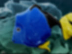
Purdue/IUPUI PEOPLE'S CHOICE
MXene Dory
Anupma Thakur, Nithin Chandran B S,
Purdue School of Eng. & Tech., IUPUI, USA
A multilayered Ti3C2Tx MXene is visualized as Dory the fish. Two-dimensional (2D) Ti3C2Tx MXene layers were formed by selective etching of Al layers from Ti3AlC2 MAX phase using hydrofluoric (HF) acid. “Whoa, whoa, whoa! Hey! Welcome to the MXene Dory's world”! The image width is 0.002 mm.
Drexel PEOPLE'S CHOICE
MXene Volcano
Umay Amara, Ruocun (John) Wang,
Drexel University, USA
Everyone has hot lava inside them in the form of love, hate, power, or greed and if it doesn't handle timely, it will take the form of a volcano and burn everything. Mother Nature also follows the same rule. So if this lava came out try to control it with cold and patience because corners can be hot red, but inside of everything is white and pure and demands the eruptions of love and peace. The image demonstrated the MXene sheet covered with ionic liquid, which is trying to intercalate inside the layers and can be seen in the form of a volcano. The image width is ~ 0.790 mm.

HONORABLE MENTION
Rainbow Twist
Marley Downes, Kyle Matthews, Veronika Sedajova,
Drexel University, USA
This image shows a MoO3/Ti3C2 hybrid electrode after cycling. Electrochemistry brings in materials from many different avenues of research, just as this image brings together two different nanomaterials into a "MXene Universe". In this image, the universe is represented on a nanoscale, showing the harmony of science. Thanks to Dr. Yury Gogotsi and Dr. Ekaterina Pomerantseva. The image width is 0.031 mm.

HONORABLE MENTION
A Broken Heart
Mahnaz Alijani, Central European Institute of Technology (CEITEC) – VUT, Czech Republic
After capturing more than 8,000 scanning electron microscopy (SEM) images (the essential characterization for my samples) of TiO2 nanotube layers, one day, I really missed my country, and suddenly, I found this broken heart while checking the SEM cross-section of my sample. An emotional sign from the materials when I was not in a good mood. The tubes have been only colored in red and a background is added on top of them. The image width is 0.005 mm.

HONORABLE MENTION
Nano Brain Signals
Borhan Aldeen Albiss, Hasan M Megdadi, Ibrahim Alkhaldi, Ahmad Malkawi, Hasan Al-Bawa'neh and Rawan Hayajneh
JUST Nanotechnology Institute, Jordan
This scanning electron microscopy image simulates the neuron signals that are being fired in your brain the moment you see this colorful image. Nanofibers and beads of polyvinylidene fluoride (PVDF) produced by electrospinning technique. Nanofibers widths are ranging between 20 and 80 nm.

HONORABLE MENTION
The Universe of Nanotechnology
Navid Keshmiri, Amir Hosein Ahmadian Hoseini, Parisa Najmi, University of British Columbia – Okanagan, Canada
This image illustrates an assembly of polystyrene (PS) beads and carbon nanotubes (CNT). The PS beads were synthesized through an emulsion-evaporation process. The PS beads and CNT were dispersed in deionized water in a bath sonicator and then vacuum-filtered. This technique was used to make a polymer nanocomposite with a segregated structure featuring high electrical conductivity for electromagnetic wave shielding applications. The image width is 0.055 mm.

HONORABLE MENTION
Dead Nano-Tree
Mahshid Mokhtarnejad,
University of Tennessee, Knoxville, TN, USA
The scanning electron micrograph shows cobalt oxide nanotrees synthesized through a simple and green technique called laser ablation synthesis in solution (LASiS) by ablating pure cobalt metal target in ethanol solution. The material can be used as a catalyst for oxygen reduction reaction (ORR). These materials are hard to synthesize with standard hydrothermal methods. The image width is 0.018 mm.
A complete list of 2022 finalists is available on NanoArtography Instagram page.
Click here to see the 2022 finalists.